
Female chest wall tumors pictures. When you have found an odd-looking, lumpy, swollen, or strange contour to the breast wall, people typically want to see some pictures before they go to see a doctor. Educational value of images. Images can help teach how these tumours may look on the surface; they should always be taken in context with clinical details and other imaging.
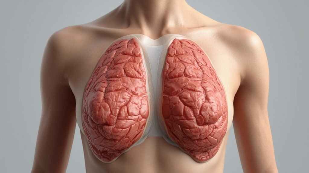
Anatomy of the Female Chest Wall
Proper interpretation of pictures of female chest wall tumors. To properly interpret images, it’s quite important to have a good grasp of anatomy beneath the skin.
According to a numbering of layers of the chest wall:
- Skin and subcutaneous fat
- Muscles (pectoralis major and minor, intercostal muscles)
- Connective tissue
- Ribs and cartilage
- Blood vessels and nerves
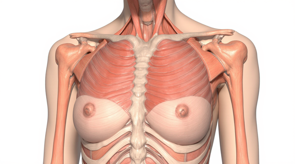
It is from these layers that tumors develop, which explains why they look so different across patients.
What Female Chest Wall Tumors Pictures Are Intended to Show
Purpose of Medical Images
Medical images of chest wall tumors are not meant to diagnose by sight alone. Instead, they demonstrate:
- External shape and contour changes
- Surface symmetry or asymmetry
- Relationship to surrounding structures
- Degree of visibility at rest
These images assist in clearer dialogue between clinicians and patients about results.
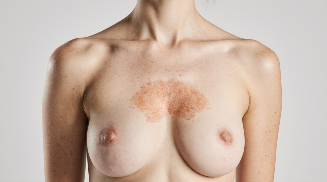
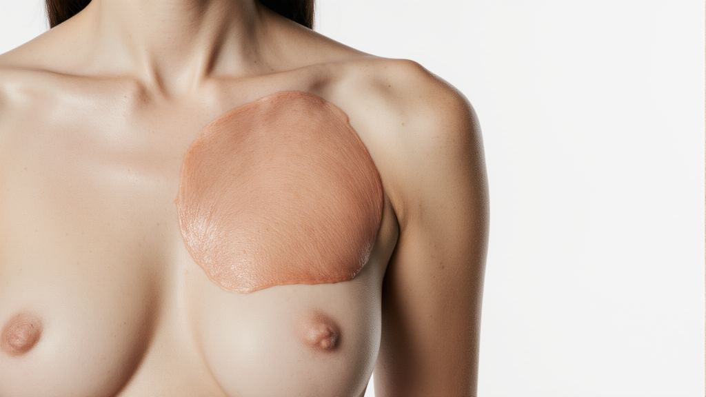
External Visual Patterns Seen in Pictures
Most female chest wall tumors pictures show one or more of the following:
- A localized bulge or protrusion
- Smooth or uneven surface contour
- Fixed or immobile appearance
- Normal or stretched skin covering the area
Of course, many skin cancers do not necessarily change the color of your skin, so the absence of redness does not mean that you are home free.
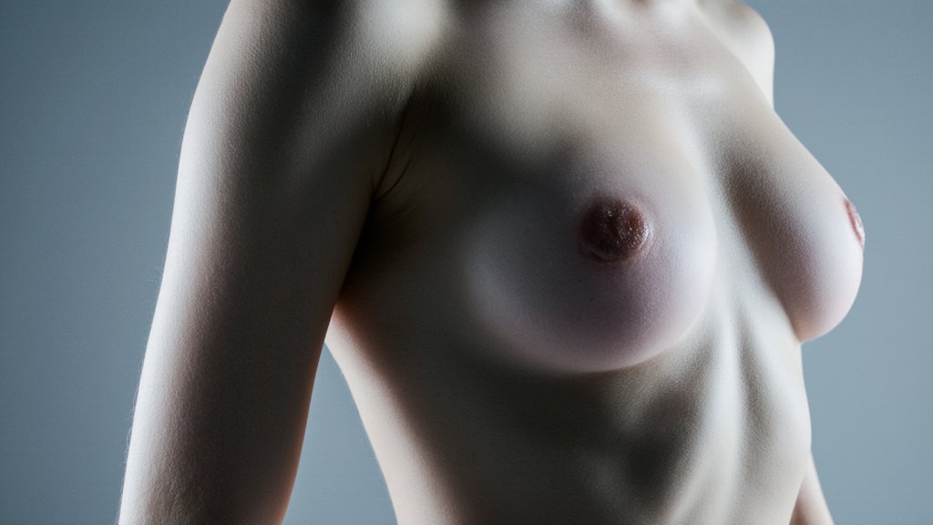
Depth and Location Influence How Tumors Appear in Pictures
Superficial Tumors
Tumors closer to the skin surface often appear as:
- Clearly defined lumps
- Rounded or oval elevations
- Soft or firm contours
These are usually easier to notice in photographs and mirrors.
Deep Chest Wall Tumors
Tumors arising from muscle, cartilage, or bone may appear as:
- Subtle asymmetry
- Gradual outward bulging
- Poorly defined edges
In pictures, these can look deceptively mild despite significant internal involvement.
Benign Findings Commonly Seen in Female Chest Wall Tumors Pictures
Typical Visual Characteristics
Benign tumors often display:
- Smooth, regular shape
- Slow or unchanged growth over time
- No skin breakdown
Examples frequently illustrated include fatty or fibrous growths that appear stable across multiple images.
Why Benign Tumors Still Appear Concerning
Even benign tumors may look alarming in pictures because:
- They distort chest symmetry
- They become more noticeable when arms are raised
- Weight loss can make them more visible
This explains why images alone often cause unnecessary anxiety.
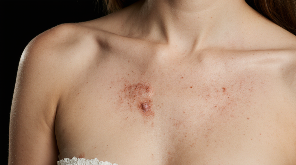
Malignant Features Sometimes Suggested in Pictures
Visual Clues Doctors Take Seriously
While pictures cannot confirm cancer, concerning visual features may include:
- Rapid size change over short periods
- Irregular or angular contours
- Skin tethering or dimpling
- Fixed position against deeper structures
These features prompt further diagnostic testing, not immediate conclusions.
Important Limitation of Visual Diagnosis
Some tumours, which are grossly dramatic and benign, may appear very subtle. This overlap makes professional evaluation essential. For broader clinical context and background information, readers can refer to a detailed chest wall tumor medical overview available through established health resources.
How Imaging Complements Female Chest Wall Tumors Pictures
Medical Imaging vs. Surface Photos
Doctors rely on internal imaging tools such as:
- X-rays for bone involvement
- CT scans for structural definition
- MRI for soft tissue clarity
Surface images reveal only what is above the skin, not the internal nature.
Why Photographs Alone Are Insufficient
Photographs cannot determine:
- Tumor origin
- Tissue composition
- Growth aggressiveness
- Cellular behavior
This is why pictures are always combined with imaging and biopsy results.
Common Symptoms That Match Visual Findings
Although pictures capture appearance, symptoms often guide urgency:
- Persistent localized pain
- Sensation of pressure or fullness
- Restricted chest movement
- Progressive enlargement
For instance, a patient may detect a painless lump in the beginning and later, several months later, pain in the breast.
Distinguishing Chest Wall Tumors from Breast Conditions in Pictures
This is a frequent source of confusion.
Chest wall tumors generally:
- Remain fixed when breast tissue moves
- Sit deeper beneath the breast
- Change appearance with posture
Breast-related lumps usually move with breast tissue and are evaluated using different imaging protocols.
Clinical Red Flags That Images May Reveal
Doctors pay attention when pictures show:
- Progressive asymmetry
- Visible enlargement across time
- Skin distortion without inflammation
- Prominent veins over the mass

These signs guide further investigation, not assumptions.
Why Self-Comparison Using Pictures Can Be Misleading
Online image searches vary widely in:
- Lighting and angles
- Body composition
- Tumor stage
- Individual anatomy
Two visually similar tumors may represent entirely different diagnoses.
What to Do After Viewing Female Chest Wall Tumors Pictures
If images raise concern, the most useful steps include:
- Documenting size and changes
- Avoiding repeated self-examination
- Scheduling a professional evaluation
- Providing a clear symptom history
Medical clarity comes from evaluation, not comparison. For more educational health-related topics and general medical awareness, readers can explore additional resources available on Lifelens Journey.
Conclusion
Female Chest Wall Tumors pictures serve an educational purpose by showing the appearance of a chest wall tumor externally. But physical ones do not necessarily indicate cause, severity, or diagnosis. Knowledge of the anatomical layers, visual constraints, and clinical world makes it possible to responsively and productively use these images.
Professional evaluation is the best way to receive a correct diagnosis and peace of mind.


One comment on “7 Important Truths About Female Chest Wall Tumors Pictures Doctors Want You to Understand”
Comments are closed.